Radiotherapy is a widely used method for treating cancer, involving the delivery of high-energy radiation beams to a defined target area from multiple directions. Accurate identification of the treatment area is critical to avoid unintended exposure to surrounding healthy tissues.
Traditionally, computed tomography (CT) has been the primary imaging modality for radiotherapy planning due to its ability to provide attenuation information needed for dose calculations. However, CT images may offer limited soft tissue contrast, therefore often requiring additional imaging modalities to support the definition of treatment volumes.
Spectronic Medical’s MRI Planner enables the use of magnetic resonance imaging (MRI) as the sole image input for radiotherapy planning. The software utilizes the deep learning-based Transfer Function Estimation algorithm to derive synthetic CT images for dose calculation from MRI data. This approach may support workflows that benefit from the soft tissue contrast of MRI.
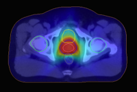
Synthetic CT with overlayed dose distribution for prostate radiotherapy
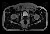
MRI of the prostate region

MRI Planner is a software solution designed to generate synthetic CT images from MRI data.

The GRADE phantom and evaluation software provides all tools required for reliable MRI scanner QA in a radiotherapy planning workflow.
Redefining radiotherapy
MRI only radiotherapy planning